
Coronaviruses which could infect both humans and animals were first identified in 1960. A total of six variants of the viruses have been identified. The current variant which is spreading all over the world, is variant 7, called the “2019 Novel Coronavirus” or “COVID-19” (originally called New Coronavirus). The World Health Organization (WHO) has declared the novel coronavirus (COVID-19) outbreak as a global pandemic. Thailand has also had a high rate of infections and deaths, causing enormous economic and social impacts. Therefore, it is imperative to stop or slow down the spread of the disease with the detection of COVID-19 in various ways.
Investigation for COVID-19 patients can be done in many ways, such as with viral genomic tests (RT-PCR), Rapid tests, Antigen Tests (RT-LAMP), etc. However, these methods are tested in a lab using limited chemicals and testing equipment, causing incomplete examinations. Therefore, Assoc. Prof. Dr. Worapan Kusakunniran, an ICT Mahidol instructor and the Head of the Mahidol Vision and Information Transfer Research LAB (MVIT), together with Prof. Dr. Thanongchai Siriapisith, Assoc. Prof. Trongtum Tongdee, Assoc. Prof. Dr. Pairash Saiviroonporn, Department of Radiology, the Faculty of Medicine Siriraj Hospital, Mr. Sarattha Karnjanapreechakorn, Miss Punyanuch Borwarnginn, and Mr. Krittanat Sutassananon, ICT Mahidol Ph.D students, developed the research entitled “COVID-19 detection and heatmap generation in chest x-ray images”.
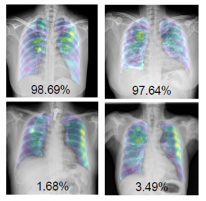
The “COVID-19 detection and heatmap generation in chest x-ray images” proposed an alternative solution, using image processing technology and deep learning to assist in automatically detecting COVID-19 from chest x-ray images. The models are trained to classify chest x-ray images into three classes including normal, COVID-19, and other viral and bacterial diseases, as well as show heatmaps with their confidence scores of being COVID-19, which can help conclude the final diagnosis.
The benefits of this research can be used to detect COVID-19 in a chest x-ray image. Furthermore, the proposed solution can be trained with other diseases in the future. Hospitals can use the software that has been developed as a device for pre-screening the patient’s chest X-ray image. In addition, it can be created as a Mobile Service to take chest x-ray images in different areas and send images to be processed in a central system. It can also be developed into a device to help guide physicians to the area where abnormalities occur, allowing them to read chest x-ray images faster and more comprehensively.
Those who are interested in reading the full article, please visit: https://www.spiedigitallibrary.org/journals/journal-of-medical-imaging/volume-8/issue-S1/014001/COVID-19-detection-and-heatmap-generation-in-chest-x-ray/10.1117/1.JMI.8.S1.014001.full?SSO=1